Urinary tract stones

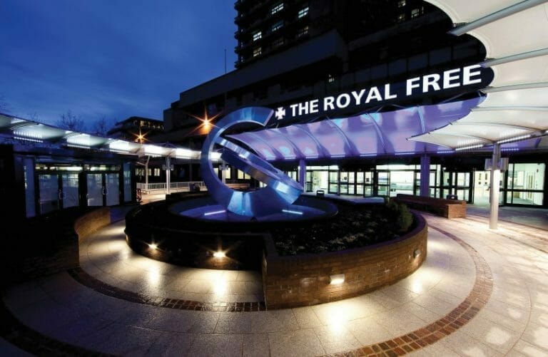
Our private Urinary tract stones services are provided at the Royal Free Hospital, Hampstead.
To find out more about our hospitals click here:
Renal stones affect approximately 1 in 10 of the population and are more common in men. They most frequently occur between the ages of 30 and 50 years and can recur even if lifestyle modifications are made.
Kidney stones are often asymptomatic and picked up during investigations for other conditions. If symptomatic they can cause back pain, blood in the urine and recurrent urinary tract infections. If the stones fall out of the kidney and block the ureter tube there is often a sudden onset of extremely severe pain. This is usually cramping in nature and centred around the loin or groin. In men it can radiate down to the testicles. Patients with ureteric colic often present to the accident and emergency department due to the severity of their symptoms.
Diagnosis is usually achieved with a non enhanced CT scan, which detects the vast majority of stone types and sizes. An ultrasound scan can also be used but it is less accurate and may miss smaller stones. It can be useful however in patient follow up to reduce radiation exposure. Plain X-rays may also detect dense stones which are normally made out of calcium but again this modality is less sensitive than CT.
If ureteric stones are small (less than 5mm) then there is a high chance that they will pass without any surgical intervention. Tablets called alpha blockers (which are usually used in men with difficulties passing urine) help to relax the ureter tube. They increase the rate of stone passage and reduce both the time to stone passage and the analgesia requirements.
If the stone fails to pass conservatively, if there is uncontrollable pain, significant blockage to the kidney, damage to kidney function or signs of infection then urgent action has to be taken. This often involves the placement of a plastic tube called a JJ stent which runs from the kidney to the bladder and unblocks the drainage system of the kidney. It is inserted under a general anaesthetic via a telescope which goes down the urethra (water pipe) and into the bladder. There are no cuts on the skin.
Further treatment to deal with renal tract stones can include:
- Extracorporeal shock wave lithotripsy (ESWL)
This is performed on an outpatient basis with the patient awake usually lying on their back. A pain killing injection or tablets are often given prior to the treatment beginning. High energy shock waves are focussed at the stone using either X ray or ultrasound to try and break it up into smaller pieces. It may take up to three treatments before it is clear if ESWL will break the stone or not. Once fragmented the pieces will be passed by the patient. Complications are uncommon but include blood in the urine, bruising around the kidney and pain as the fragments pass or become stuck in the ureter. - Ureteroscopy
This is performed under a general anaesthetic usually as a day case procedure. A small telescope is placed via the urethra into the bladder and then up into the ureter and kidney if necessary. The stone is seen directly, often broken up with a laser and the fragments removed with a basket. A JJ stent is usually inserted at the end of the operation to stop the kidney becoming blocked by swelling of the ureter tube, blood or stone fragments. Major complications are rare with damage to the ureter being the most feared. - Percutaneous nephrolithotomy (PCNL)
This is usually reserved for the treatment of larger stones in the kidney or upper ureter. It involves being admitted to the hospital and staying in for several days. Under a general anaesthetic a small incision is made in the skin and the kidney punctured using a fine needle. This tract is then dilated and a plastic tube inserted into the kidney through which a telescope can be passed. The stone is then identified and either removed whole or broken into small pieces. After the operation it is usual to be left with a small tube coming from the kidney for several days. Complications include: failure to access the kidney, bleeding and damage to surrounding organs.
Once the stone is removed the main aim of treatment is to prevent recurrence. Lifestyle measures such as maintaining a high fluid intake (2-3l/day) and reducing the amount of salt and animal protein in the diet can be useful. Exact instructions will depend on the type of stone that is formed.
The Royal Free Private Patients Unit has an experienced team of stone surgeons who offer the highest quality care. We are one of the only units in London to perform PCNLs in the supine position. This can reduce anaesthetic problems and make access to the kidney easier from the bladder.
We also have a consultant nephrologist as part of our team who can investigate the specific cause of the stones, as well as other medical issues that can affect kidney function. From this, an individualised management plan can be created in order to reduce the chance of further stone formation.
Get in touch
